COGNITOIAS · BIOLOGY MASTER NOTES · FINAL V4
HUMAN DIGESTIVE SYSTEM & DIGESTIVE ENZYMES
The Food Journey · Think–Predict–Discover · Very Easy English · UPSC / UPPSC / State PCS + selected NEET/AIPMT concepts
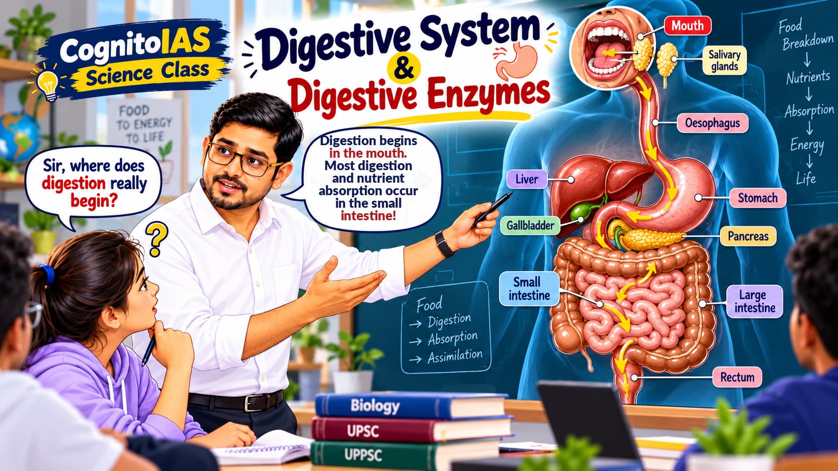
Imagine your lunch: roti + dal + a little ghee. You swallow it in seconds. But how does this food reach your cells?
Roti brings starch. Dal brings proteins. Ghee brings fat. All three take the same road, but need different enzymes.
The mystery: Where does each start breaking down? Who supplies the enzymes? Where do the small nutrients finally go?
How to read this lesson
At each stop: PREDICT the answer → DISCOVER the reason → BEAT THE EXAM TRAP. Try the small checkpoints before looking at the answer. No memorisation without understanding!
Quick Navigation
What must food become before your cells can use it?
1. First Understand the Big Idea
DISCOVERY DIALOGUE · GUESS FIRST, THEN READ
Food travels through one continuous tube
This tube is the alimentary canal / gastrointestinal (GI) tract. The small intestine has three parts: Duodenum → Jejunum → Ileum.
Who helps from outside?
Salivary glands produce saliva.
Liver produces bile.
Gallbladder stores and concentrates bile.
Pancreas produces pancreatic juice.
START HERE
2. Digestion, Absorption and Assimilation Are Different
Ingestion: taking food into the mouth.
Digestion: breaking complex food into smaller molecules (for example, starch → smaller sugars).
Absorption: nutrients cross the intestinal lining into blood or lymph.
Assimilation: body cells use the absorbed nutrients (for example, glucose supplies energy).
Egestion: removal of undigested residue as faeces through the anus.
EXAM TRAP: Egestion ≠ Excretion
3. Two Types of Digestion
Mechanical digestion: physical breakdown—chewing in mouth and churning in stomach.
Chemical digestion: enzymes break chemical bonds in large molecules, for example starch —amylase→ smaller sugars.
RECALL
A teacher crushes a biscuit but uses no enzyme. Which kind of digestion happened?
What happens to the roti while you chew?
4. Mouth — Digestion Starts Here
DISCOVERY DIALOGUE · GUESS FIRST, THEN READ
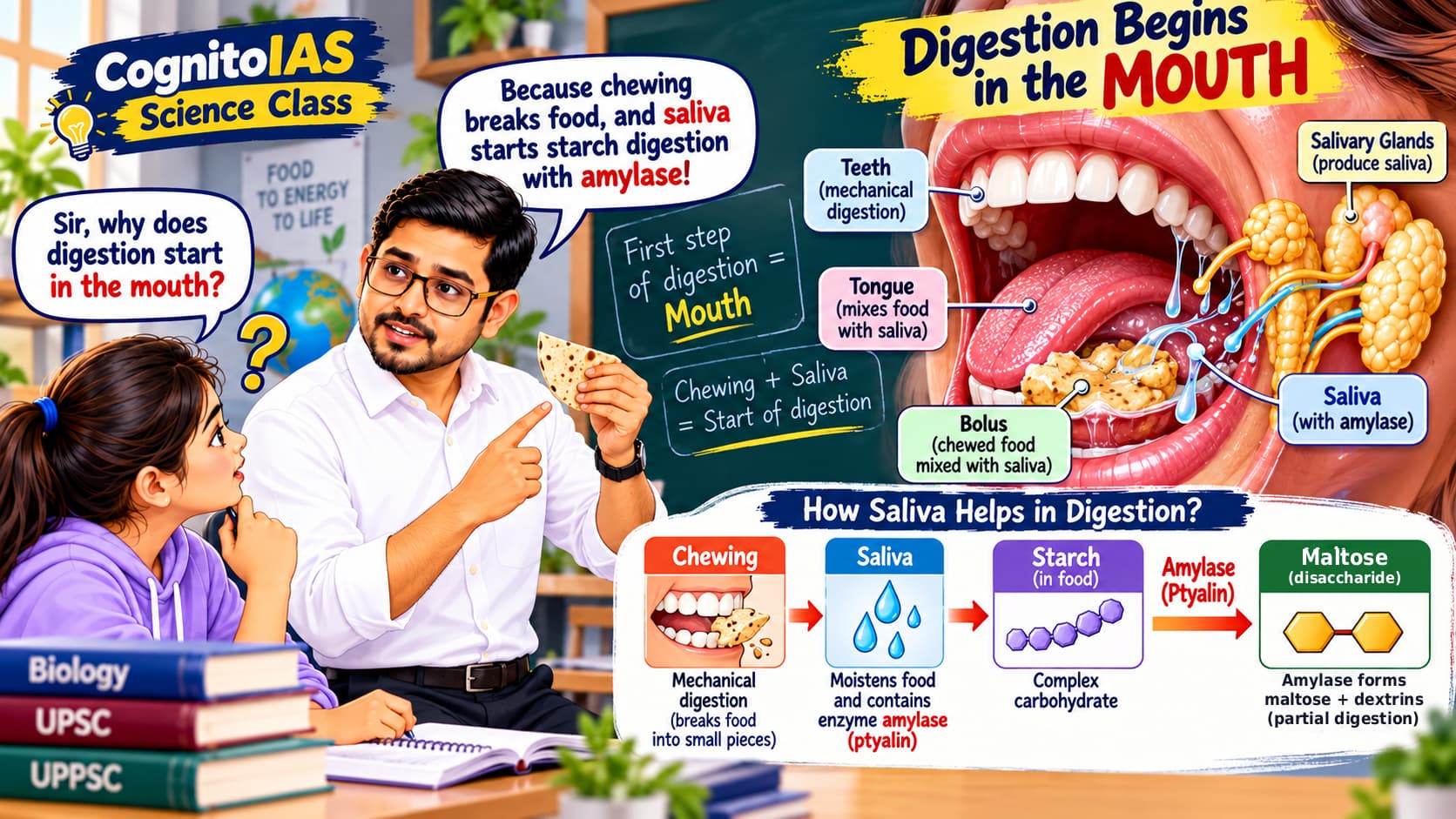
Three pairs of major salivary glands
Parotid
Submandibular / Submaxillary
Sublingual
Saliva helps swallowing and contains protective substances. Its main starch-digesting enzyme is salivary amylase (ptyalin).
EXAM FACT
5. Does Saliva Digest Protein?
6. What Happens During Swallowing?
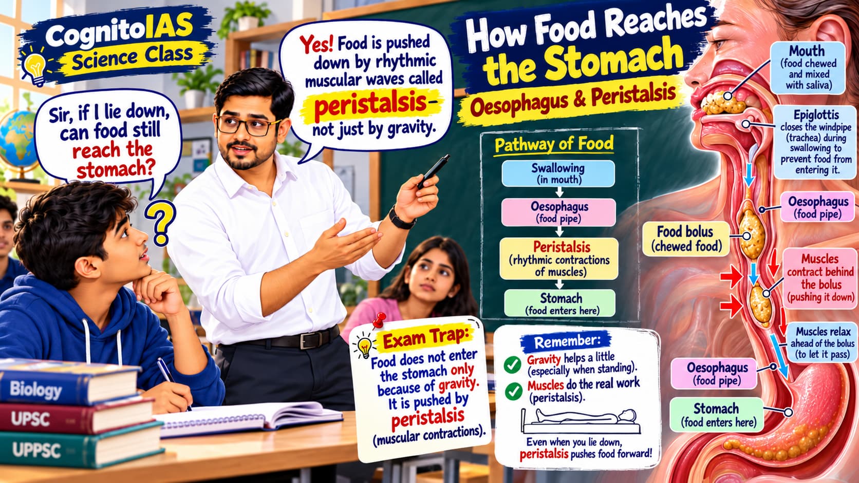
Food passes from mouth → pharynx → oesophagus. During swallowing, the airway is protected by coordinated movements, including the epiglottis, which helps cover the entrance to the windpipe.
EXAM FACT
Food accidentally starts entering the airway. Name the protective flap and one reflex.
7. Oesophagus — Does Gravity Pull Food Down?
DISCOVERY DIALOGUE · GUESS FIRST, THEN READ
Peristalsis also helps move contents through other parts of the GI tract. The oesophagus is mainly a transport tube, not a major site of chemical digestion.
REMEMBER
What begins to happen to dal proteins?
8. Stomach — The Mixing Chamber
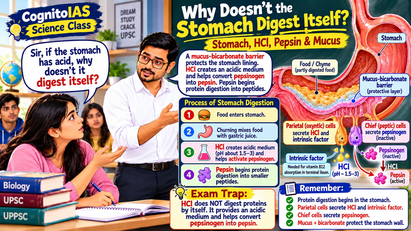
The semi-fluid mixture leaving the stomach is called chyme. Food passes towards the duodenum through the pyloric sphincter.
9. Hydrochloric Acid — Why Is Acid in the Stomach?
DISCOVERY DIALOGUE · GUESS FIRST, THEN READ
HCl is secreted by parietal (oxyntic) cells.
It creates an acidic medium for gastric protein digestion.
It helps pepsinogen → pepsin.
It helps limit many microbes.
NEET / AIPMT CELL FACT
10. Why Doesn't the Stomach Digest Itself?
DISCOVERY DIALOGUE · GUESS FIRST, THEN READ
Mucus–bicarbonate barrier protects the surface.
Tight epithelial junctions limit acid entry.
The lining can repair and renew itself.
Chief cells first release inactive pepsinogen, not large amounts of active pepsin.
CELL MAP — STOMACH
Parietal / oxyntic cells → HCl + intrinsic factor
Surface mucous cells → protective gastric mucus.
11. Pepsin — Protein Digestion Begins Here
Stomach chief cells release pepsinogen. Acidic conditions activate it into pepsin; pepsin can also help activate more pepsinogen.
SCIENTIFIC CORRECTION
GOLDEN RULE
True or false: pepsin converts a whole protein directly into free amino acids.
12. Pepsin, Trypsin and Chymotrypsin — Do Not Confuse
Pepsin: stomach enzyme; acts in acidic stomach.
Trypsin: released by pancreas as inactive trypsinogen; acts in small intestine.
Chymotrypsin: released by pancreas as inactive chymotrypsinogen; acts in small intestine.
EXAM TRAP
Food does not enter the liver or pancreas. Their juices enter the food tube.
13. Liver — The Largest Gland
The liver is the largest gland in the human body. Its major digestive secretion is bile. It also processes absorbed nutrients, synthesises many plasma proteins, helps detoxify substances, and converts toxic ammonia into urea.
EXAM LINK
14. Gallbladder — Does It Produce Bile?
Liver = Factory: produces bile.
Gallbladder = Storage tank: stores and concentrates bile.
Duodenum = Delivery point: receives bile through bile ducts.
MEMORY
15. Bile — Is It a Digestive Enzyme?
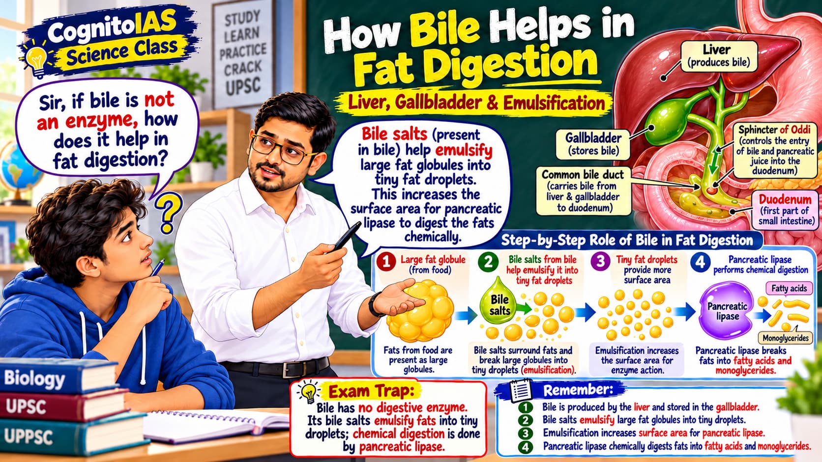
DISCOVERY DIALOGUE · GUESS FIRST, THEN READ
Emulsification disperses one large fat globule into many tiny droplets, increasing surface area for pancreatic lipase.
WRITE PRECISELY
Choose the correct pair: (A) Bile = enzyme, (B) Bile = emulsifier.
16. Pancreas — One Organ, Two Jobs
Exocrine part: sends enzyme-rich pancreatic juice into duodenum.
Endocrine part: releases hormones such as insulin and glucagon into blood.
Detailed insulin/glucagon actions belong to the separate Endocrine System notes.
17. Why Is Pancreatic Juice So Important?
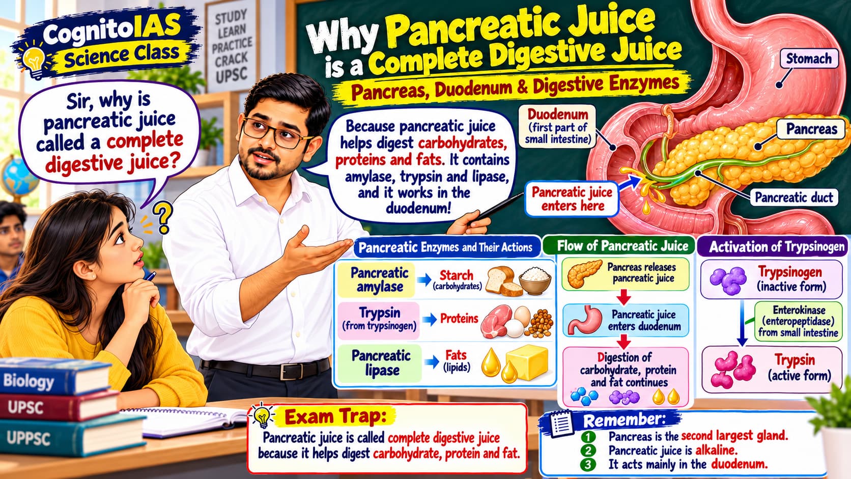
Think of the pancreas as a multi-tool kit: one tool works on starch, another on proteins, another on fat, and another on DNA/RNA. Bicarbonate prepares a suitable working environment. Now learn each tool by name.
Pancreatic juice enters the duodenum and is rich in digestive enzymes and bicarbonate.
Pancreatic amylase: starch → smaller sugars.
Trypsin, chymotrypsin, carboxypeptidase: proteins / peptides → smaller peptides and amino-acid products.
Pancreatic lipase: triglycerides → mainly fatty acids + monoglycerides.
Pancreatic nucleases: DNA/RNA → nucleotides.
Bicarbonate: neutralises acidic chyme, helping pancreatic enzymes work.
OLDER EXAM PHRASE
CAUTION
18. Why Doesn't the Pancreas Digest Itself?
DISCOVERY DIALOGUE · GUESS FIRST, THEN READ
Enteropeptidase (enterokinase) is located on the duodenal brush border. Once active, trypsin helps activate other pancreatic zymogens.
PANCREATIC ZYMOGEN MAP
Chymotrypsinogen → Chymotrypsin
Procarboxypeptidase → Carboxypeptidase.
A question says: pancreatic trypsinogen becomes active in the stomach. Correct it.
The busiest station: finish digestion, then absorb nutrients.
19. Small Intestine — The Main Digestion and Absorption Centre
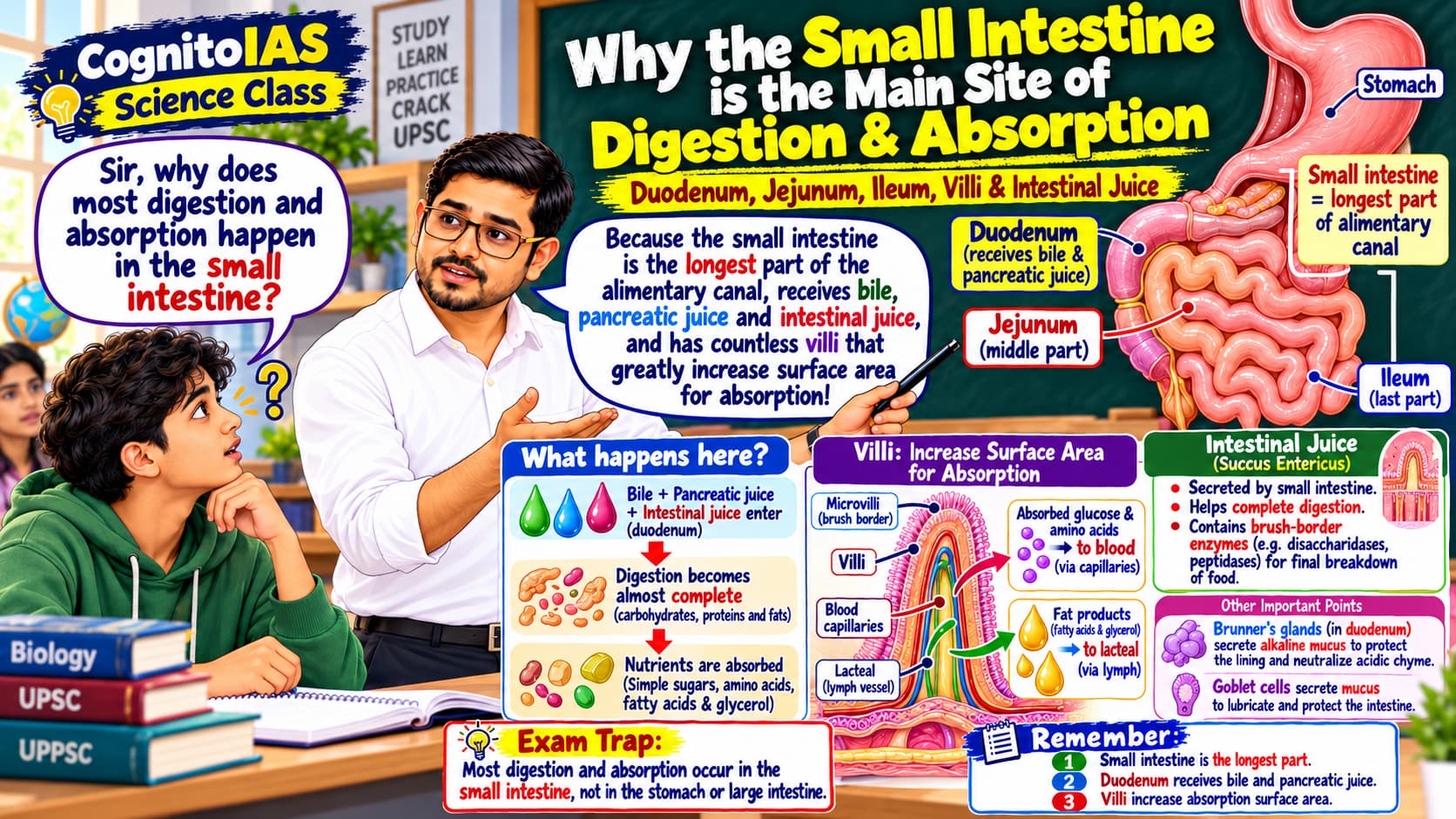
DISCOVERY DIALOGUE · GUESS FIRST, THEN READ
It is the longest part of the alimentary canal, though narrower than the large intestine. It is highly coiled and has three parts:
EXAM FACT
20. Duodenum — The Mixing Junction
DISCOVERY DIALOGUE · GUESS FIRST, THEN READ
The duodenum is the first and shortest part of the small intestine. It receives acidic chyme from stomach, bile from liver/gallbladder, and pancreatic juice from pancreas.
Acid is neutralised.
Fat is emulsified and acted on by lipase.
Carbohydrates and proteins undergo further enzyme digestion.
NEET/AIPMT BOOSTER
Brunner’s glands → duodenal submucosa → bicarbonate-rich alkaline mucus that helps protect against acid.
21. Why Must Stomach Acid Be Neutralised?
Protects the duodenal lining.
Creates a better pH for pancreatic enzymes.
Helps inactivate stomach pepsin after food moves onward.
22. Jejunum and Ileum — Major Absorption Continues
Duodenum participates in both digestion and absorption. The jejunum is a major nutrient-absorption site, and the ileum continues absorption.
Jejunum: much carbohydrate and protein absorption.
Terminal ileum: especially important for bile salts and vitamin B12–intrinsic factor complex.
Ileum opens into caecum at the ileo-caecal junction.
OLD IAS LENGTH ORDER
Which region is especially important for intrinsic factor–vitamin B12 uptake?
23. Why Is the Small Intestine So Good at Absorption?
DISCOVERY DIALOGUE · GUESS FIRST, THEN READ
Villi are finger-like projections of the intestinal lining. Microvilli are tiny projections on the surface of absorptive epithelial cells.
EXAM FACT
24. What Goes Into Blood and What Goes Into Lymph?
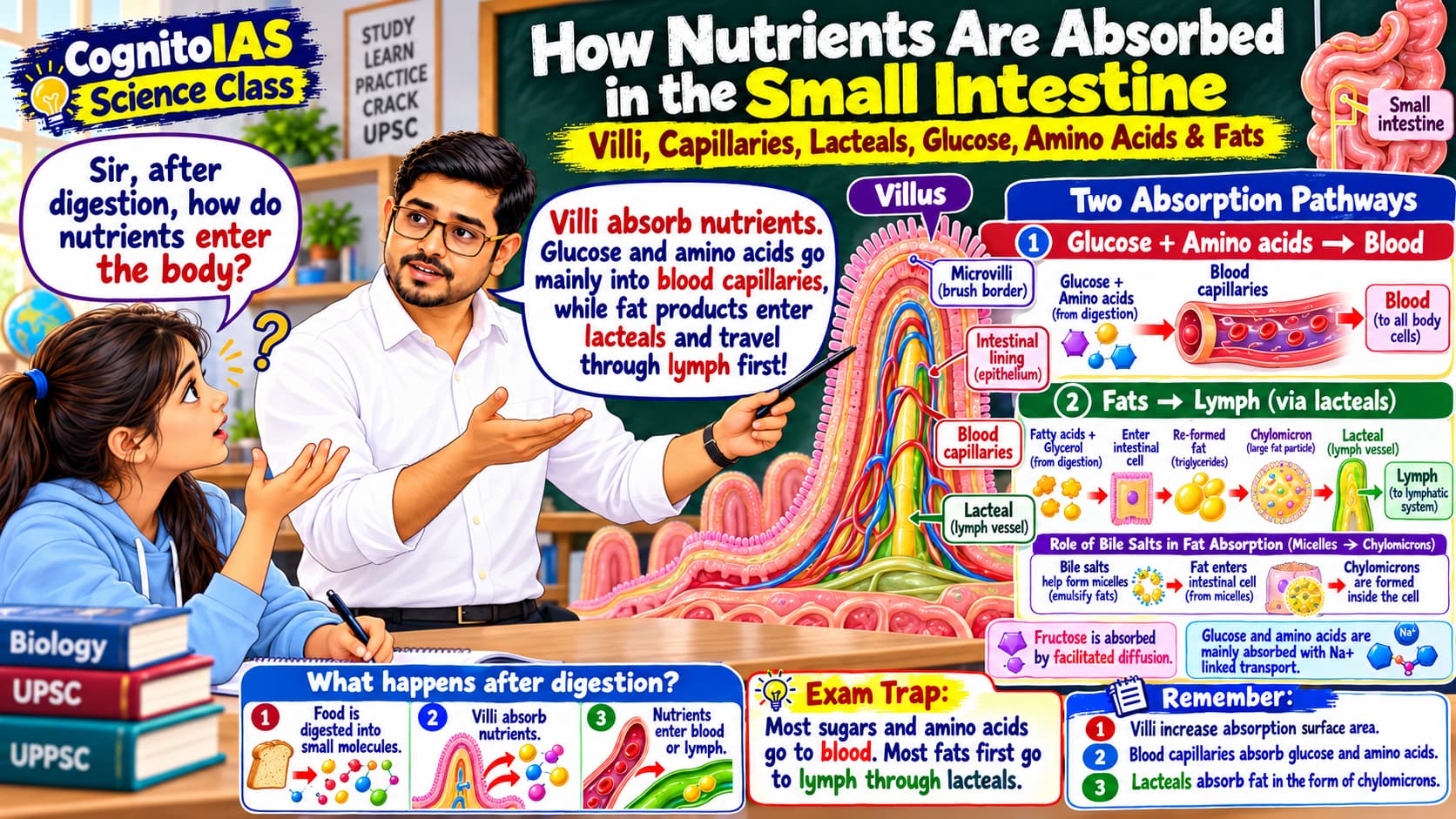
Two travel routes: water-friendly nutrients mostly use the blood route. Most long-chain fats need extra packing and first take the lymph route. Read the two routes below.
DISCOVERY DIALOGUE · GUESS FIRST, THEN READ
Water-soluble nutrients
Glucose and galactose: absorbed by Na⁺-linked co-transport into intestinal cells.
Fructose: absorbed mainly by facilitated diffusion.
Many amino acids: absorbed by carrier-mediated transport, often Na⁺-linked.
These then pass into blood capillaries and reach the liver through the portal circulation.
Most long-chain dietary fats
Micelles formed with bile salts carry fatty acids and monoglycerides to the cell surface. Micelles themselves do not normally enter the intestinal cells intact; their lipid contents enter. The cells reassemble triglycerides and package them as chylomicrons.
EXAM MEMORY
Fructose → facilitated diffusion
Most long-chain fats → micelles → chylomicrons → lacteals → lymph → blood.
SMALL EXCEPTION
Fill the route: Fat product → ___ → intestinal cell → ___ → lacteal.
25. What Is Chyle?
DO NOT CONFUSE
Chyle = milky, fat-rich intestinal lymph.
26. Intestinal Enzymes — Final Finishing Work
The finishing desk: amylase does not finish starch digestion all alone. Brush-border enzymes make the small absorbable sugars.
DISCOVERY DIALOGUE · GUESS FIRST, THEN READ
The intestinal mucosa produces intestinal juice (succus entericus). Many enzymes that finish digestion are attached to the brush border of intestinal cells.
Maltase: maltose → 2 glucose.
Sucrase: sucrose → glucose + fructose.
Lactase: lactose → glucose + galactose.
Peptidases: smaller peptides → amino acids.
Nucleotidases / nucleosidases / phosphatases: act on products of nucleic-acid digestion.
NEET / AIPMT TERMS
STARCH DIGESTION SEQUENCE
NEET / AIPMT Booster — Cells and Glands
Goblet cells → Mucus (lubrication + protection).
Paneth cells → Lysozyme and other antimicrobial substances; found in intestinal crypts (crypts of Lieberkühn).
Chief/peptic cells → Pepsinogen.
Parietal/oxyntic cells → HCl + intrinsic factor.
Hepatocytes / hepatic cells → Bile.
Brunner’s glands → Duodenal alkaline mucus.
SIX-LINE PYQ CELL MAP
Amylase has already acted on starch. Which enzyme can finish maltose into glucose?
27. What Are the Final Absorbable Products?
Remember the goal: digestion is successful only when food has become small enough for intestinal cells to absorb.
Carbohydrates → Monosaccharides: glucose, fructose, galactose.
Proteins → mainly amino acids (some di-/tripeptides are also taken into intestinal cells and then further broken down).
Triglycerides → mainly free fatty acids + monoglycerides; old elementary books simplify this as fatty acids + glycerol.
Nucleic acids → smaller components such as nitrogenous bases, pentose sugars and phosphate.
EXAM TRAP
What happens to the leftovers after nutrients are absorbed?
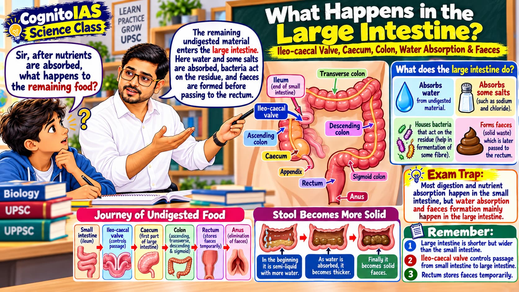
28. Large Intestine — Why Is It Needed?
DISCOVERY DIALOGUE · GUESS FIRST, THEN READ
Caecum begins the large intestine.
Colon reclaims remaining water and salts.
Rectum stores faeces before egestion.
It is not the major site of enzymatic digestion. Some dietary fibre is fermented by gut microbes.
PYQ CELL FACT
29. Why Is the Small Intestine Called “Small”?
REMEMBER
Large intestine = shorter, wider.
True or false: the large intestine is longer than the small intestine.
30. Appendix — Where Is It Attached?
The vermiform appendix is a blind-ended tube attached to the caecum, at the beginning of the large intestine.
VALVE NEARBY
31. Why Can't Humans Digest Cellulose?
DISCOVERY DIALOGUE · GUESS FIRST, THEN READ
Cellulose therefore largely behaves as dietary fibre. Some fibre components can be fermented by intestinal microbes, but our own enzymes do not digest cellulose like starch.
EXAM TRAP
Distinguish hormones, enzymes and older textbook expressions.
32. Hormone or Enzyme? — A Major Exam Trap
DISCOVERY DIALOGUE · GUESS FIRST, THEN READ
Gastrin: hormone; stimulates gastric acid secretion and stomach activity.
Secretin: hormone released mainly by duodenum in response to acidic chyme; promotes bicarbonate-rich pancreatic secretion.
Cholecystokinin (CCK): hormone released mainly in response to fats/proteins; stimulates gallbladder contraction and enzyme-rich pancreatic secretion.
Pepsin, trypsin, amylase and lipase: digestive enzymes.
EXAM MEMORY
Pepsin + Trypsin + Amylase + Lipase = ENZYMES.
The options are pepsin, trypsin, ptyalin and gastrin. Which one is not an enzyme?
33. Enterogastrone — An Older Exam Term
Enterogastrone is an older umbrella term for intestinal hormonal effects that reduce gastric secretion or emptying, especially when fat reaches the small intestine. Modern physiology discusses individual hormones instead of treating enterogastrone as one major, precisely defined hormone.
OLD IAS-STYLE FACT
Fat in small intestine → inhibitory intestinal hormonal responses (older term: enterogastrone).
34. Renin vs Rennin — Do Not Confuse
Renin (one n after e): enzyme released by kidney; acts on angiotensinogen in blood-pressure regulation. Not a digestive enzyme.
Rennin / chymosin (two n after e): milk-clotting protease classically described in young mammals. Older exam books sometimes associate it with infant gastric juice.
OLD AIPMT CAUTION
Your exam says “renin digests milk.” Spot the spelling and concept trap.
35. Rapid Recall — Bile vs Lipase
EXAM TRAP
36. Rapid Recall — Where Does Each Food Start Digestion?
Carbohydrate: begins in mouth — salivary amylase.
Protein: begins in stomach — pepsin.
Fat: some digestion can occur through lingual/gastric lipases, but the bulk occurs in small intestine through pancreatic lipase.
Nucleic acids: mainly digested in small intestine by pancreatic nucleases and intestinal finishing enzymes.
OLD AIPMT CAUTION
See the complete enzyme map, traps and one-minute journey.
37. Digestive Enzyme Master Map
Revision task: cover the enzyme names with your hand. Look at the food and location, then say the enzyme before checking.
Mouth
Salivary amylase / ptyalin: starch → maltose and dextrins.
Stomach
Pepsin: proteins → smaller peptides.
Gastric lipase: limited fat digestion.
Pancreas (acts in small intestine)
Pancreatic amylase: starch → smaller carbohydrates.
Trypsin / chymotrypsin: proteins → peptides.
Carboxypeptidase: releases amino acids from peptide ends.
Pancreatic lipase: triglycerides → fatty acids + monoglycerides.
Nucleases: DNA/RNA → nucleotides.
Small-intestinal brush border
Maltase: maltose → glucose.
Sucrase: sucrose → glucose + fructose.
Lactase: lactose → glucose + galactose.
Peptidases: small peptides → amino acids.
Nucleotidases / nucleosidases: finish nucleic-acid digestion.
FAST ENZYME RECALL
38. What If Lactase Is Missing?
DISCOVERY DIALOGUE · GUESS FIRST, THEN READ
Lactose is the major sugar in milk. It is broken down by the intestinal brush-border enzyme lactase into glucose and galactose.
DO NOT CONFUSE
39. Gallstones — Why Can They Affect Fat Digestion?
DISCOVERY DIALOGUE · GUESS FIRST, THEN READ
EXAM LINK
A blocked bile duct mainly affects the digestion of starch, protein or fat?
40. Common Exam Traps — Old + New
The original 15 traps are retained, and PYQ-driven additions have been appended:
✓ It begins in mouth.
✓ Most chemical digestion occurs in small intestine.
✓ It begins in stomach.
✓ Liver produces; gallbladder stores.
✓ Bile contains no digestive enzyme.
✓ Bile emulsifies; lipase chemically digests.
✓ Gastrin is a hormone.
✓ It comes from pancreas and acts in small intestine.
✓ Most long-chain fats enter lacteals → lymph first.
✓ It is attached to caecum.
✓ Humans lack cellulase.
✓ They are different processes.
✓ Chyme = stomach mixture; chyle = milky lymph.
✓ Renin is linked to kidney/RAAS.
✓ It is an older exam expression for pancreatic juice.
✓ It means intestinal juice.
✓ It regulates the hepatopancreatic opening in duodenum.
✓ It helps prevent backflow.
✓ Goblet cells secrete mucus.
✓ They secrete antimicrobial substances including lysozyme.
✓ They are in duodenum.
✓ Fructose uses facilitated diffusion.
✓ Micelles deliver lipid contents to the cell surface.
✓ Pepsin mainly produces peptides.
✓ Limited lingual/gastric lipase activity can occur earlier.
41. One-Minute Journey — Tell the Story Yourself
Chewing + saliva → starch digestion begins
Peristalsis transports food
HCl + pepsin → protein digestion begins; chyme forms
Bile + pancreatic juice → major chemical digestion
Major absorption into blood and lymph
Mainly water + electrolyte absorption; faeces form
Stores faeces
Egestion
OUR LUNCH, FINALLY EXPLAINED
Roti (starch): mouth amylase starts; pancreatic amylase continues; brush-border enzymes finish sugars; glucose enters blood.
Dal (proteins): stomach pepsin starts; pancreatic proteases and intestinal peptidases finish; amino acids enter blood.
Ghee (fat): limited early lipase action is possible; bile emulsifies; pancreatic lipase breaks fat; lipid products reach cells in micelles; most long-chain lipids leave as chylomicrons through lacteals.
Now answer without looking: Why does our body need more than one enzyme?
42. Final Memory Box — Revision in 90 Seconds
Say aloud these six answers before reading the check-line:
Where does starch digestion begin, protein digestion begin, and most fat digestion occur?
Who produces bile? Who stores it?
Which intestinal enzyme turns trypsinogen into trypsin?
Why does the small intestine have villi?
How does fructose absorption differ from glucose absorption?
How do most long-chain fats reach the blood?
If you can explain WHY, not only WHAT, the concept is yours.
COGNITOIAS • Digestive System Final V3 • English • CKEditor / Mobile / Full Width
COGNITOIAS · बायोलॉजी मास्टर नोट्स · हिंदी-हिंग्लिश V4
मानव पाचन तंत्र (Human Digestive System) और पाचक एंजाइम (Digestive Enzymes)
भोजन की यात्रा · सोचिए–अनुमान लगाइए–समझिए · सरल हिंदी + English scientific terms · यूपीएससी / यूपीपीएससी / राज्य पीसीएस
हमारा मिशन: भोजन के एक कौर की यात्रा
ये नोट्स कैसे पढ़ें?
झटपट नेविगेशन
कोशिकाएँ भोजन का उपयोग करने से पहले उसमें क्या बदलता है?
1. सबसे पहले पाचन की बड़ी बात समझें
खोजपूर्ण संवाद · पहले सोचें, फिर समझें
भोजन किस लगातार नली से गुजरता है?
इसी रास्ते को आहार नाल (Alimentary Canal) या जठरांत्र मार्ग (Gastrointestinal Tract / GI Tract) कहते हैं। छोटी आँत के तीन भाग हैं: ग्रहणी (Duodenum) → मध्यांत्र (Jejunum) → शेषांत्र (Ileum)।
बाहर से मदद कौन करता है?
लार ग्रंथियाँ (Salivary Glands) → लार (Saliva) बनाती हैं।
यकृत (Liver) → पित्त (Bile) बनाता है।
पित्ताशय (Gallbladder) → पित्त को जमा और गाढ़ा करता है।
अग्न्याशय (Pancreas) → अग्न्याशयी रस (Pancreatic Juice) बनाता है।
शुरुआती सूत्र
2. पाचन, अवशोषण और स्वांगीकरण में अंतर
खोजपूर्ण संवाद · पहले सोचें, फिर समझें
अंतर्ग्रहण (Ingestion): भोजन को मुँह में लेना।
पाचन (Digestion): बड़े अणुओं को छोटे अणुओं में तोड़ना; जैसे स्टार्च से छोटी शर्कराएँ।
अवशोषण (Absorption): पोषक तत्वों का आँत की दीवार पार करके रक्त (Blood) या लसीका (Lymph) में पहुँचना।
स्वांगीकरण (Assimilation): कोशिकाओं द्वारा अवशोषित पदार्थों का उपयोग; जैसे ग्लूकोज़ से ऊर्जा मिलना।
मलत्याग (Egestion): अपचित पदार्थ का मल (Faeces) के रूप में गुदा से बाहर निकलना।
परीक्षा में भ्रम: मलत्याग ≠ उत्सर्जन
3. पाचन के दो प्रकार
खोजपूर्ण संवाद · पहले सोचें, फिर समझें
यांत्रिक पाचन (Mechanical Digestion): चबाना और आमाशय में मथना; भोजन भौतिक रूप से छोटे टुकड़ों में टूटता है।
रासायनिक पाचन (Chemical Digestion): एंजाइम (Enzymes) रासायनिक बंध तोड़ते हैं; जैसे एमाइलेज (Amylase) स्टार्च को छोटी शर्कराओं में बदलता है।
याद रखें
शिक्षक ने बिस्किट को केवल कुचल दिया, कोई एंजाइम नहीं लगाया। यह कौन-सा पाचन है?
चबाते समय रोटी में क्या होता है?
4. मुख (Mouth) — पाचन की शुरुआत यहीं
खोजपूर्ण संवाद · पहले सोचें, फिर समझें
तीन जोड़ी प्रमुख लार ग्रंथियाँ
कर्णमूल ग्रंथि (Parotid Gland)
अधोहनु ग्रंथि (Submandibular / Submaxillary Gland)
अधोजिह्वा ग्रंथि (Sublingual Gland)
लार (Saliva) निगलने में मदद करती है और उसमें सुरक्षा देने वाले पदार्थ भी होते हैं। इसका प्रमुख स्टार्च-पाचक एंजाइम लार एमाइलेज (Salivary Amylase / Ptyalin) है।
परीक्षा तथ्य
वैज्ञानिक सावधानी
5. क्या लार प्रोटीन भी पचाती है?
खोजपूर्ण संवाद · पहले सोचें, फिर समझें
6. निगलते समय क्या होता है?
भोजन मुख (Mouth) → ग्रसनी (Pharynx) → ग्रासनली (Oesophagus) से गुजरता है। निगलने के दौरान कई समन्वित क्रियाएँ श्वसन मार्ग की रक्षा करती हैं। कंठनाल ढक्कन (Epiglottis) श्वासनली के प्रवेश द्वार को ढकने में मदद करता है।
खोजपूर्ण संवाद · पहले सोचें, फिर समझें
परीक्षा तथ्य
भोजन गलती से वायुमार्ग में जाने लगे तो सुरक्षात्मक ढक्कन और एक प्रतिवर्त बताइए।
7. ग्रासनली (Oesophagus) — क्या गुरुत्वाकर्षण भोजन नीचे खींचता है?
खोजपूर्ण संवाद · पहले सोचें, फिर समझें
क्रमाकुंचन (Peristalsis) जठरांत्र मार्ग (GI Tract) के अन्य हिस्सों में भी पदार्थों को आगे बढ़ाता है। ग्रासनली रासायनिक पाचन (Chemical Digestion) का प्रमुख स्थान नहीं है।
याद रखें
दाल के प्रोटीन में क्या बदलाव आता है?
8. आमाशय (Stomach) — भोजन को मिलाने और मथने वाला कक्ष
चित्र पढ़ते समय ध्यान दें
खोजपूर्ण संवाद · पहले सोचें, फिर समझें
आमाशय से निकलने वाला अर्द्धतरल भोजन काइम (Chyme) कहलाता है। भोजन जठरनिर्गम संवरणी (Pyloric Sphincter) से होकर ग्रहणी (Duodenum) में जाता है।
9. हाइड्रोक्लोरिक अम्ल — आमाशय में अम्ल क्यों?
खोजपूर्ण संवाद · पहले सोचें, फिर समझें
HCl का स्राव पार्श्विका कोशिकाएँ (Parietal Cells) करती हैं।
यह आमाशय में अम्लीय माध्यम (Acidic Medium) बनाता है।
यह पेप्सिनोजन → पेप्सिन (Pepsinogen → Pepsin) सक्रिय होने में मदद करता है।
यह कई सूक्ष्मजीवों को नियंत्रित करने में भी सहायक है।
नीट / एआईपीएमटी कोशिका तथ्य
10. आमाशय खुद को क्यों नहीं पचा लेता?
खोजपूर्ण संवाद · पहले सोचें, फिर समझें
श्लेष्मा–बाइकार्बोनेट अवरोध (Mucus–Bicarbonate Barrier) सतह की रक्षा करता है।
उपकला की सघन संधियाँ (Tight Epithelial Junctions) अम्ल को भीतर जाने से रोकती हैं।
आमाशय की परत जल्दी मरम्मत और नवीनीकरण कर सकती है।
मुख्य / पेप्टिक कोशिकाएँ (Chief / Peptic Cells) पहले निष्क्रिय पेप्सिनोजन (Pepsinogen) छोड़ती हैं।
आमाशय की कोशिकाओं का नक्शा
11. पेप्सिन — प्रोटीन पाचन की शुरुआत
आमाशय की मुख्य कोशिकाएँ (Chief Cells) पेप्सिनोजन (Pepsinogen) बनाती हैं। अम्लीय वातावरण उसे सक्रिय पेप्सिन (Pepsin) में बदलने में मदद करता है। सक्रिय पेप्सिन और पेप्सिनोजन को भी सक्रिय कर सकता है।
वैज्ञानिक सावधानी
गोल्डन रूल
सही या गलत: पेप्सिन पूरे प्रोटीन को सीधे मुक्त अमीनो अम्ल में बदल देता है।
पुराने पीवाईक्यू (PYQ) की भाषा
12. पेप्सिन, ट्रिप्सिन और काइमोट्रिप्सिन में अंतर
खोजपूर्ण संवाद · पहले सोचें, फिर समझें
पेप्सिन (Pepsin): आमाशय का एंजाइम; अम्लीय आमाशय में काम करता है।
ट्रिप्सिन (Trypsin): अग्न्याशय से निष्क्रिय ट्रिप्सिनोजन (Trypsinogen) के रूप में निकलता है; छोटी आँत में काम करता है।
काइमोट्रिप्सिन (Chymotrypsin): अग्न्याशय से निष्क्रिय काइमोट्रिप्सिनोजन (Chymotrypsinogen) के रूप में निकलता है; छोटी आँत में काम करता है।
परीक्षा में भ्रम
भोजन इनके अंदर से नहीं जाता; इनके रस आहार नाल में पहुँचते हैं।
13. यकृत (Liver) — शरीर की सबसे बड़ी ग्रंथि
यकृत (Liver) मानव शरीर की सबसे बड़ी ग्रंथि (Largest Gland) है। पाचन के लिए इसका मुख्य स्राव पित्त (Bile) है। यह अवशोषित पोषक तत्वों की प्रक्रिया करता है, कई प्लाज़्मा प्रोटीन (Plasma Proteins) बनाता है, हानिकारक पदार्थों को कम करने में मदद करता है और विषैले अमोनिया (Ammonia) को यूरिया (Urea) में बदलता है।
खोजपूर्ण संवाद · पहले सोचें, फिर समझें
परीक्षा लिंक
14. पित्ताशय (Gallbladder) — क्या यह पित्त बनाता है?
खोजपूर्ण संवाद · पहले सोचें, फिर समझें
यकृत (Liver) = फैक्ट्री: पित्त (Bile) बनाता है।
पित्ताशय (Gallbladder) = टंकी: पित्त को रखता और गाढ़ा करता है।
ग्रहणी (Duodenum) = पहुँचने का स्थान: पित्त नलिकाओं (Bile Ducts) से पित्त प्राप्त करती है।
याद करने का सूत्र
15. पित्त (Bile) — क्या यह पाचक एंजाइम है?
खोजपूर्ण संवाद · पहले सोचें, फिर समझें
पायसीकरण (Emulsification) में बड़ी वसा-बूँद कई छोटी बूँदों में फैलती है। इससे लाइपेज के लिए सतही क्षेत्रफल (Surface Area) बढ़ जाता है।
सटीक लिखें
सही जोड़ी चुनें: (A) पित्त = पाचक एंजाइम, (B) पित्त = पायसीकारक (Emulsifier)।
16. अग्न्याशय (Pancreas) — एक अंग, दो काम
खोजपूर्ण संवाद · पहले सोचें, फिर समझें
बहिःस्रावी भाग (Exocrine Part): एंजाइमयुक्त अग्न्याशयी रस (Pancreatic Juice) ग्रहणी (Duodenum) में भेजता है।
अंतःस्रावी भाग (Endocrine Part): इंसुलिन (Insulin) और ग्लूकागॉन (Glucagon) जैसे हार्मोन रक्त में छोड़ता है।
इंसुलिन और ग्लूकागॉन के विस्तृत काम अलग अंतःस्रावी तंत्र (Endocrine System) के notes में पढ़ेंगे।
17. अग्न्याशयी रस (Pancreatic Juice) इतना महत्वपूर्ण क्यों है?
एंजाइम सक्रियता (Enzyme Activation)
अग्न्याशय को कई औज़ारों वाला टूलकिट समझिए—एक स्टार्च पर, दूसरा प्रोटीन पर, तीसरा वसा पर और चौथा डीएनए/आरएनए (DNA/RNA) पर काम करता है। बाइकार्बोनेट (Bicarbonate) इन एंजाइमों के लिए उपयुक्त माध्यम बनाता है।
अग्न्याशयी रस (Pancreatic Juice) ग्रहणी में पहुँचता है और पाचक एंजाइमों के साथ बाइकार्बोनेट (Bicarbonate) से समृद्ध होता है।
अग्न्याशयी एमाइलेज (Pancreatic Amylase): स्टार्च → छोटी शर्कराएँ।
ट्रिप्सिन (Trypsin), काइमोट्रिप्सिन (Chymotrypsin), कार्बोक्सीपेप्टिडेज़ (Carboxypeptidase): प्रोटीन/पेप्टाइड → छोटे पेप्टाइड और अमीनो-अम्ल वाले उत्पाद।
अग्न्याशयी लाइपेज (Pancreatic Lipase): ट्राइग्लिसराइड (Triglycerides) → मुख्यतः वसीय अम्ल (Fatty Acids) + मोनोग्लिसराइड (Monoglycerides)।
अग्न्याशयी न्यूक्लिएज़ (Pancreatic Nucleases): डीएनए/आरएनए (DNA/RNA) → न्यूक्लियोटाइड (Nucleotides)।
बाइकार्बोनेट (Bicarbonate): अम्लीय काइम (Acidic Chyme) को उदासीन कर एंजाइमों की सहायता करता है।
पुरानी परीक्षा की भाषा
सावधानी
18. अग्न्याशय खुद को क्यों नहीं पचा देता?
खोजपूर्ण संवाद · पहले सोचें, फिर समझें
एंटेरोपेप्टिडेज़ (Enteropeptidase / Enterokinase) ग्रहणी की ब्रश बॉर्डर (Brush Border) सतह पर होता है। सक्रिय ट्रिप्सिन आगे दूसरे ज़ाइमोजन (Zymogens) को सक्रिय करता है।
अग्न्याशयी ज़ाइमोजन मानचित्र
एक प्रश्न कहता है: अग्न्याशयी ट्रिप्सिनोजन आमाशय में सक्रिय होता है। इसे सही कीजिए।
यहाँ रोटी, दाल और घी का अंतिम पाचन कैसे होता है?
19. छोटी आँत (Small Intestine) — पाचन और अवशोषण का मुख्य केंद्र
चित्र की वैज्ञानिक स्पष्टता
खोजपूर्ण संवाद · पहले सोचें, फिर समझें
छोटी आँत लंबी और अत्यधिक कुंडलित (Highly Coiled) होती है। इसके भाग हैं—ग्रहणी (Duodenum) → मध्यांत्र (Jejunum) → शेषांत्र (Ileum)।
परीक्षा तथ्य
20. ग्रहणी (Duodenum) — सभी पाचक रसों का मिलन स्थल
खोजपूर्ण संवाद · पहले सोचें, फिर समझें
ग्रहणी (Duodenum) छोटी आँत का पहला और सबसे छोटा भाग है। इसे आमाशय से अम्लीय काइम, यकृत/पित्ताशय से पित्त और अग्न्याशय से अग्न्याशयी रस मिलता है।
अम्ल का काफी भाग उदासीन (Neutralised) होता है।
वसा का पायसीकरण (Emulsification) होता है और लाइपेज काम करता है।
कार्बोहाइड्रेट और प्रोटीन का आगे एंजाइमी पाचन होता है।
नीट / एआईपीएमटी बूस्टर
21. आमाशय का अम्ल उदासीन करना क्यों जरूरी है?
खोजपूर्ण संवाद · पहले सोचें, फिर समझें
यह ग्रहणी (Duodenum) की दीवार की रक्षा करता है।
यह अग्न्याशयी एंजाइमों (Pancreatic Enzymes) के लिए बेहतर पीएच (pH) बनाता है।
भोजन आगे बढ़ने पर आमाशय के पेप्सिन (Pepsin) को निष्क्रिय होने में सहायता मिलती है।
22. मध्यांत्र (Jejunum) और शेषांत्र (Ileum) — अवशोषण जारी रहता है
ग्रहणी (Duodenum) पाचन और अवशोषण दोनों में भाग लेती है। मध्यांत्र (Jejunum) पोषक अवशोषण का प्रमुख स्थान है और शेषांत्र (Ileum) आगे अवशोषण जारी रखता है।
मध्यांत्र (Jejunum): अनेक कार्बोहाइड्रेट तथा प्रोटीन-पाचन उत्पादों का अवशोषण।
अंतिम शेषांत्र (Terminal Ileum): पित्त लवण (Bile Salts) और विटामिन बी-12–अंतर्जात कारक संकुल (Vitamin B12–Intrinsic Factor Complex) के अवशोषण के लिए खास महत्त्वपूर्ण।
शेषांत्र (Ileum), सीकम (Caecum) से शेषांत्र–सीकम संधि (Ileo-caecal Junction) पर जुड़ता है।
पुराना IAS लंबाई क्रम
अंतर्जात कारक से बँधे विटामिन बी-12 का अवशोषण विशेष रूप से कहाँ होता है?
23. छोटी आँत इतने अधिक पोषक तत्व कैसे सोखती है?
खोजपूर्ण संवाद · पहले सोचें, फिर समझें
विलस (Villi) आँत की अंदरूनी परत की उँगली-जैसी उभरी संरचनाएँ हैं। सूक्ष्मविलस (Microvilli) अवशोषक उपकला कोशिकाओं (Absorptive Epithelial Cells) की सतह पर बहुत छोटे उभार हैं।
परीक्षा तथ्य
24. कौन-से पोषक तत्व रक्त में और कौन-से लसीका में?
रक्त बनाम लसीका — सही तीर देखिए
पानी में आसानी से घुलने वाले पोषक तत्व सामान्यतः रक्त मार्ग (Blood Route) से जाते हैं। लंबी श्रृंखला वाली अधिकांश वसा को विशेष पैकिंग चाहिए, इसलिए वह पहले लसीका मार्ग (Lymph Route) अपनाती है।
खोजपूर्ण संवाद · पहले सोचें, फिर समझें
जल-विलेय पोषक तत्वों का प्रवेश
ग्लूकोज़ (Glucose) और गैलेक्टोज़ (Galactose): सोडियम-संबद्ध सहपरिवहन (Na⁺-linked Co-transport) से आँत की कोशिका में।
फ्रक्टोज़ (Fructose): मुख्यतः सुगमित विसरण (Facilitated Diffusion) से।
अनेक अमीनो अम्ल (Amino Acids): वाहक-मध्यस्थित परिवहन (Carrier-mediated Transport), अक्सर Na⁺-संबद्ध।
फिर ये रक्त केशिकाओं → पोर्टल परिसंचरण (Portal Circulation) → यकृत तक पहुँचते हैं।
लंबी श्रृंखला वाली अधिकांश वसा का रास्ता
पित्त लवण (Bile Salts) से बने माइसेल वसीय अम्ल (Fatty Acids) और मोनोग्लिसराइड (Monoglycerides) को कोशिका तक लाते हैं। माइसेल स्वयं सामान्यतः पूरा भीतर नहीं जाता। कोशिका ट्राइग्लिसराइड (Triglycerides) फिर बनाती है और उन्हें काइलोमाइक्रॉन (Chylomicrons) में पैक करती है।
परीक्षा स्मृति
छोटा अपवाद
रास्ता पूरा करें: वसा-पाचन उत्पाद → ____ → आँत की कोशिका → ____ → लैक्टियल।
एक लाइन में: ग्लूकोज़ और गैलेक्टोज़ का प्रवेश सोडियम-संबद्ध है; फ्रक्टोज़ सुगमित विसरण से आता है; कई अमीनो अम्ल भी सोडियम-संबद्ध वाहक इस्तेमाल करते हैं। अधिकांश लंबी श्रृंखला वाली वसा पहले लसीका में जाती है। सभी पोषक तत्वों का मार्ग एक-जैसा नहीं है।
25. काइल (Chyle) क्या है?
खोजपूर्ण संवाद · पहले सोचें, फिर समझें
भ्रम न करें
26. आँत के एंजाइम — पाचन का अंतिम काम
अंतिम पाचन को फिनिशिंग डेस्क समझिए। अकेला एमाइलेज स्टार्च को पूरी तरह नहीं पचाता। ब्रश बॉर्डर एंजाइम (Brush-border Enzymes) अवशोषित होने योग्य छोटी शर्कराएँ बनाते हैं।
खोजपूर्ण संवाद · पहले सोचें, फिर समझें
आँत्रिक श्लेष्मकला (Intestinal Mucosa) आँत्र रस (Succus Entericus) बनाती है। अंतिम पाचन पूरा करने वाले बहुत-से एंजाइम ब्रश बॉर्डर (Brush Border) से जुड़े रहते हैं।
माल्टेज़ (Maltase): माल्टोज़ (Maltose) → 2 ग्लूकोज़ (Glucose)।
सुक्रेज़ (Sucrase): सुक्रोज़ (Sucrose) → ग्लूकोज़ + फ्रक्टोज़।
लैक्टेज़ (Lactase): लैक्टोज़ (Lactose) → ग्लूकोज़ + गैलेक्टोज़।
पेप्टिडेज़ (Peptidases): छोटे पेप्टाइड → अमीनो अम्ल (Amino Acids)।
न्यूक्लियोटिडेज़ / न्यूक्लियोसिडेज़ / फॉस्फेटेज़ (Nucleotidases / Nucleosidases / Phosphatases): न्यूक्लिक अम्ल पाचन के उत्पादों पर काम करते हैं।
नीट / एआईपीएमटी की भाषा
स्टार्च पाचन का क्रम
नीट / एआईपीएमटी: कोशिकाएँ और ग्रंथियाँ
गॉब्लेट कोशिकाएँ (Goblet Cells) → श्लेष्मा (Mucus); चिकनाई और सुरक्षा।
पैनेथ कोशिकाएँ (Paneth Cells) → लाइसोज़ाइम (Lysozyme) और अन्य रोगाणुरोधी पदार्थ; आँत्र क्रिप्ट / लाइबरकुन की क्रिप्ट (Crypts of Lieberkühn) में पाई जाती हैं।
मुख्य / पेप्टिक कोशिकाएँ (Chief / Peptic Cells) → पेप्सिनोजन (Pepsinogen)।
पार्श्विका / ऑक्सिंटिक कोशिकाएँ (Parietal / Oxyntic Cells) → HCl + अंतर्जात कारक (Intrinsic Factor)।
यकृत कोशिकाएँ / हेपेटोसाइट (Hepatocytes / Hepatic Cells) → पित्त (Bile)।
ब्रूनर ग्रंथियाँ (Brunner’s Glands) → ग्रहणी का क्षारीय श्लेष्मा (Duodenal Alkaline Mucus)।
छह लाइन का पीवाईक्यू सेल-मैप
एमाइलेज ने स्टार्च पर काम कर दिया। माल्टोज़ को ग्लूकोज़ में कौन-सा एंजाइम बदलता है?
27. पाचन के अंतिम अवशोषित होने योग्य उत्पाद
याद रखें: पाचन तभी उपयोगी है जब भोजन इतने छोटे अणुओं में बदल जाए कि आँत की कोशिकाएँ उन्हें अवशोषित (Absorb) कर सकें।
कार्बोहाइड्रेट (Carbohydrates) → मोनोसैकेराइड (Monosaccharides): ग्लूकोज़ (Glucose), फ्रक्टोज़ (Fructose), गैलेक्टोज़ (Galactose)।
प्रोटीन (Proteins) → मुख्यतः अमीनो अम्ल (Amino Acids): कुछ डाइपेप्टाइड/ट्राइपेप्टाइड (Di-/Tripeptides) भी कोशिका में जाकर आगे टूट सकते हैं।
ट्राइग्लिसराइड (Triglycerides) → मुख्यतः मुक्त वसीय अम्ल (Free Fatty Acids) + मोनोग्लिसराइड (Monoglycerides): पुरानी प्रारंभिक पुस्तकों में “वसीय अम्ल + ग्लिसरॉल” सरल रूप में मिलता है।
न्यूक्लिक अम्ल (Nucleic Acids) → नाइट्रोजन क्षारक (Nitrogenous Bases), पेंटोज़ शर्करा (Pentose Sugars) और फॉस्फेट (Phosphate) जैसे छोटे घटक।
परीक्षा में भ्रम
पोषक तत्व सोखने के बाद बचे पदार्थ का क्या होता है?
28. बड़ी आँत (Large Intestine) — इसकी जरूरत क्यों है?
पानी के अवशोषण पर सावधानी
खोजपूर्ण संवाद · पहले सोचें, फिर समझें
सीकम (Caecum) से बड़ी आँत की शुरुआत होती है।
बृहदान्त्र / कोलन (Colon) शेष पानी और लवण वापस सोखता है।
मलाशय (Rectum) मलत्याग से पहले मल जमा रखता है।
बड़ी आँत एंजाइमी पाचन (Enzymatic Digestion) का मुख्य केंद्र नहीं है।
कुछ आहारीय रेशे (Dietary Fibre) को आँत के सूक्ष्मजीव किण्वित करते हैं।
पीवाईक्यू कोशिका तथ्य
29. लंबी होते हुए भी छोटी आँत को “छोटी” क्यों कहते हैं?
खोजपूर्ण संवाद · पहले सोचें, फिर समझें
याद रखें
सही या गलत: बड़ी आँत छोटी आँत से अधिक लंबी है।
30. अपेंडिक्स (Appendix) कहाँ जुड़ा होता है?
वर्मीफॉर्म अपेंडिक्स (Vermiform Appendix) एक बंद सिरे वाली पतली नली है, जो बड़ी आँत की शुरुआत में सीकम (Caecum) से जुड़ी रहती है।
खोजपूर्ण संवाद · पहले सोचें, फिर समझें
पास का वाल्व
31. मनुष्य सेल्यूलोज़ क्यों नहीं पचा सकता?
खोजपूर्ण संवाद · पहले सोचें, फिर समझें
सेल्यूलोज़ (Cellulose) मुख्यतः आहारीय रेशा (Dietary Fibre) जैसा व्यवहार करता है। कुछ रेशों को सूक्ष्मजीव आंशिक रूप से किण्वित कर सकते हैं, लेकिन हमारे अपने एंजाइम सेल्यूलोज़ को स्टार्च जैसा नहीं पचाते।
परीक्षा में भ्रम
एंजाइम, हार्मोन और पुराने परीक्षा शब्दों में फर्क कीजिए।
32. हार्मोन या एंजाइम — एक जरूरी परीक्षा-ट्रैप
खोजपूर्ण संवाद · पहले सोचें, फिर समझें
गैस्ट्रिन (Gastrin): हार्मोन; आमाशय के अम्ल स्राव और गतिविधि को बढ़ाता है।
सीक्रेटिन (Secretin): हार्मोन; अम्लीय काइम आने पर मुख्यतः ग्रहणी से निकलता है और बाइकार्बोनेट-समृद्ध अग्न्याशयी स्राव बढ़ाता है।
कोलिसिस्टोकाइनिन (Cholecystokinin / CCK): हार्मोन; वसा/प्रोटीन आने पर निकलता है, पित्ताशय संकुचन और एंजाइम-समृद्ध अग्न्याशयी स्राव बढ़ाता है।
पेप्सिन (Pepsin), ट्रिप्सिन (Trypsin), एमाइलेज (Amylase), लाइपेज (Lipase): पाचक एंजाइम (Digestive Enzymes)।
परीक्षा स्मृति
विकल्प: पेप्सिन, ट्रिप्सिन, टायलिन और गैस्ट्रिन। इनमें कौन एंजाइम नहीं है?
33. एंटेरोगैस्ट्रोन (Enterogastrone) — पुराना परीक्षा शब्द
एंटेरोगैस्ट्रोन (Enterogastrone) एक पुराना सामूहिक नाम है। इसका उपयोग आँत के उन हार्मोन-संबंधी प्रभावों के लिए किया जाता था, जो आमाशय के स्राव या खाली होने की गति को कम करते हैं—विशेषकर जब वसा छोटी आँत तक पहुँचती है।
आधुनिक शरीर-विज्ञान (Modern Physiology) में किसी एक पूर्णतः परिभाषित प्रमुख हार्मोन के रूप में “एंटेरोगैस्ट्रोन” पर निर्भर रहने के बजाय अलग-अलग आँत्र हार्मोन पढ़े जाते हैं।
पुराना IAS शैली तथ्य
34. रेनिन (Renin) बनाम रेन्निन (Rennin) / काइमोसिन (Chymosin)
इन English spellings को जरूर अलग पहचानिए:
रेनिन (Renin; एक ‘n’ बाद में): वृक्क / किडनी (Kidney) से निकलने वाला एंजाइम; रक्तचाप नियंत्रण में एंजियोटेंसिनोजन (Angiotensinogen) पर काम करता है। यह पाचक एंजाइम नहीं है।
रेन्निन / काइमोसिन (Rennin / Chymosin; ‘nn’): दूध जमाने वाला प्रोटीएज़ (Milk-clotting Protease), जिसे छोटे स्तनधारियों के संदर्भ में पढ़ाया जाता है। पुरानी पुस्तकों में इसे शिशु जठर रस से जोड़ा मिलता है।
पुराना एआईपीएमटी सावधानी
प्रश्न कहता है: “Renin दूध पचाता है।” इसमें spelling और concept का भ्रम बताइए।
35. झटपट दोहराएँ — पित्त बनाम लाइपेज
खोजपूर्ण संवाद · पहले सोचें, फिर समझें
परीक्षा में भ्रम
36. हर खाद्य पदार्थ का पाचन कहाँ शुरू होता है?
कार्बोहाइड्रेट (Carbohydrates): मुँह में शुरू — लार एमाइलेज (Salivary Amylase)।
प्रोटीन (Proteins): आमाशय में शुरू — पेप्सिन (Pepsin)।
वसा (Fats): जिह्वीय/जठरीय लाइपेज (Lingual / Gastric Lipases) से कुछ शुरुआती पाचन संभव है, लेकिन मुख्य भाग छोटी आँत में अग्न्याशयी लाइपेज से पचता है।
न्यूक्लिक अम्ल (Nucleic Acids): मुख्यतः छोटी आँत में अग्न्याशयी न्यूक्लिएज़ (Pancreatic Nucleases) और आँत के अंतिम एंजाइमों से।
पुराना एआईपीएमटी सावधानी
अब एंजाइम मानचित्र, परीक्षा भ्रम और पूरी भोजन-यात्रा को जोड़िए।
37. पाचक एंजाइमों का पूरा मास्टर मैप
रिविज़न अभ्यास: नीचे एंजाइम के नाम हाथ से ढकिए। केवल भोजन और स्थान देखकर एंजाइम बोलिए, फिर जाँचिए।
मुख (Mouth)
लार एमाइलेज / टायलिन (Salivary Amylase / Ptyalin): स्टार्च → माल्टोज़ + डेक्स्ट्रिन।
आमाशय (Stomach)
पेप्सिन (Pepsin): प्रोटीन → छोटे पेप्टाइड।
जठरीय लाइपेज (Gastric Lipase): वसा का सीमित पाचन।
अग्न्याशय से, काम छोटी आँत में (Pancreatic Enzymes)
अग्न्याशयी एमाइलेज (Pancreatic Amylase): स्टार्च → छोटे कार्बोहाइड्रेट।
ट्रिप्सिन / काइमोट्रिप्सिन (Trypsin / Chymotrypsin): प्रोटीन → पेप्टाइड।
कार्बोक्सीपेप्टिडेज़ (Carboxypeptidase): पेप्टाइड के सिरों से अमीनो अम्ल अलग करता है।
अग्न्याशयी लाइपेज (Pancreatic Lipase): ट्राइग्लिसराइड → वसीय अम्ल + मोनोग्लिसराइड।
न्यूक्लिएज़ (Nucleases): डीएनए/आरएनए → न्यूक्लियोटाइड।
छोटी आँत का ब्रश बॉर्डर (Small Intestinal Brush Border)
माल्टेज़ (Maltase): माल्टोज़ → ग्लूकोज़।
सुक्रेज़ (Sucrase): सुक्रोज़ → ग्लूकोज़ + फ्रक्टोज़।
लैक्टेज़ (Lactase): लैक्टोज़ → ग्लूकोज़ + गैलेक्टोज़।
पेप्टिडेज़ (Peptidases): छोटे पेप्टाइड → अमीनो अम्ल।
न्यूक्लियोटिडेज़ / न्यूक्लियोसिडेज़ (Nucleotidases / Nucleosidases): न्यूक्लिक अम्ल का अंतिम पाचन।
एंजाइम की तेज़ याद
38. लैक्टेज़ कम हो तो क्या होता है?
खोजपूर्ण संवाद · पहले सोचें, फिर समझें
दूध की प्रमुख शर्करा लैक्टोज़ (Lactose) है। आँत के ब्रश बॉर्डर का लैक्टेज़ (Lactase) इसे ग्लूकोज़ (Glucose) + गैलेक्टोज़ (Galactose) में तोड़ता है।
भ्रम न करें
39. पित्त-पथरी से वसा पाचन प्रभावित क्यों हो सकता है?
खोजपूर्ण संवाद · पहले सोचें, फिर समझें
परीक्षा लिंक
पित्त नलिका बंद होने पर स्टार्च, प्रोटीन या वसा—किसके पाचन में मुख्य समस्या होगी?
40. परीक्षा के 25 सामान्य भ्रम — पुराने और नए
पुराने 15 और नए पीवाईक्यू (PYQ) आधारित कुल 25 आम भ्रम:
✗ पाचन आमाशय से शुरू होता है।
✗ अधिकांश पाचन आमाशय में होता है।
✗ प्रोटीन पाचन छोटी आँत में शुरू होता है।
✗ पित्ताशय पित्त बनाता है।
✗ पित्त एक पाचक एंजाइम है।
✗ पित्त वसा का रासायनिक पाचन करता है।
✗ गैस्ट्रिन एक एंजाइम है।
✗ ट्रिप्सिन आमाशय का एंजाइम है।
✗ सभी पोषक पदार्थ सीधे रक्त में जाते हैं।
✗ अपेंडिक्स छोटी आँत से जुड़ा है।
✗ मनुष्य एमाइलेज से सेल्यूलोज़ पचाता है।
✗ मलत्याग और उत्सर्जन एक ही हैं।
✗ काइम और काइल एक हैं।
✗ रेनिन (Renin) पाचक एंजाइम है।
✗ “संपूर्ण पाचक रस” आधुनिक औपचारिक वर्गीकरण है।
✗ सक्सस एंटेरिकस (Succus Entericus) जठर रस है।
✗ ऑड्डी संवरणी शेषांत्र–सीकम जोड़ पर है।
✗ इलियो-सीकल वाल्व पदार्थ को इलियम में उल्टा जाने देता है।
✗ गॉब्लेट कोशिकाएँ HCl बनाती हैं।
✗ पैनेथ कोशिकाएँ पेप्सिन बनाती हैं।
✗ ब्रूनर ग्रंथियाँ आमाशय में होती हैं।
✗ फ्रक्टोज़ भी ग्लूकोज़ जैसे Na⁺ सहपरिवहन से जाता है।
✗ पूरा माइसेल आँत की कोशिका के अंदर चला जाता है।
✗ पेप्सिन सीधे पूरा प्रोटीन अमीनो अम्ल बना देता है।
✗ वसा का सारा पाचन केवल छोटी आँत में शुरू होता है।
41. एक मिनट में पूरी भोजन-यात्रा खुद सुनाइए
मुख (Mouth)
ग्रासनली (Oesophagus)
आमाशय (Stomach)
ग्रहणी (Duodenum)
मध्यांत्र + शेषांत्र (Jejunum + Ileum)
बड़ी आँत (Large Intestine)
मलाशय (Rectum)
गुदा (Anus)
हमारे भोजन का पूरा हिसाब
रोटी (Starch): मुँह का एमाइलेज शुरू करता है; अग्न्याशयी एमाइलेज आगे बढ़ाता है; ब्रश बॉर्डर एंजाइम अंतिम शर्करा बनाते हैं; ग्लूकोज़ रक्त में जाता है।
दाल (Proteins): आमाशय का पेप्सिन शुरू करता है; अग्न्याशयी प्रोटीएज़ और आँत के पेप्टिडेज़ पूरा करते हैं; अमीनो अम्ल रक्त में जाते हैं।
घी (Fat): सीमित शुरुआती लाइपेज क्रिया संभव है; पित्त पायसीकरण करता है; अग्न्याशयी लाइपेज वसा तोड़ता है; माइसेल पदार्थ कोशिका तक लाते हैं; अधिकांश लंबी श्रृंखला वाली वसा काइलोमाइक्रॉन बनकर लैक्टियल से जाती है।
अब बिना देखे बताइए: शरीर को एक से अधिक पाचक एंजाइम क्यों चाहिए?
42. अंतिम स्मृति बॉक्स — 90 सेकंड में पुनरावृत्ति
पाचन तंत्र (Digestive System) = आहार नाल + सहायक अंग।
मुख (Mouth): एमाइलेज (Amylase / Ptyalin) → स्टार्च।
आमाशय (Stomach): HCl + पेप्सिन (Pepsin) → प्रोटीन पाचन शुरू।
चीफ़ कोशिकाएँ (Chief Cells) → पेप्सिनोजन; पार्श्विका (Parietal Cells) → HCl + अंतर्जात कारक (Intrinsic Factor)।
यकृत (Liver): सबसे बड़ी ग्रंथि; पित्त, यूरिया निर्माण, ग्लाइकोजन भंडारण।
पित्ताशय (Gallbladder): पित्त जमा करता और गाढ़ा करता है।
पित्त (Bile): कोई पाचक एंजाइम नहीं; वसा का पायसीकरण करता है।
अग्न्याशय (Pancreas): मिश्रित ग्रंथि (Mixed Gland); बहिःस्रावी रस + अंतःस्रावी हार्मोन।
अग्न्याशयी रस (Pancreatic Juice): एमाइलेज + प्रोटीएज़ + लाइपेज + न्यूक्लिएज़ + बाइकार्बोनेट।
एंटेरोपेप्टिडेज़ (Enteropeptidase) → ट्रिप्सिनोजन को ट्रिप्सिन बनाता है।
छोटी आँत (Small Intestine): सबसे लंबी; अधिकांश पाचन और अवशोषण।
ग्रहणी (Duodenum): पित्त + अग्न्याशयी रस; ऑड्डी संवरणी (Sphincter of Oddi) नलिका के खुलाव पर।
ब्रूनर ग्रंथियाँ (Brunner’s Glands): ग्रहणी का क्षारीय श्लेष्मा।
सक्सस एंटेरिकस (Succus Entericus): आँत्र रस।
विलस + सूक्ष्मविलस (Villi + Microvilli): अवशोषण क्षेत्र बढ़ाते हैं।
गॉब्लेट (Goblet) → श्लेष्मा; पैनेथ (Paneth) → लाइसोज़ाइम।
ग्लूकोज़/गैलेक्टोज़: Na⁺ सहपरिवहन; फ्रक्टोज़: सुगमित विसरण।
माइसेल (Micelles) → एंटेरोसाइट (Enterocyte) में वसा → काइलोमाइक्रॉन (Chylomicrons) → लैक्टियल (Lacteals) → लसीका → रक्त।
काइम (Chyme): आमाशय मिश्रण; काइल (Chyle): दूधिया लसीका।
बड़ी आँत (Large Intestine): शेष पानी और इलेक्ट्रोलाइट का अवशोषण।
इलियो-सीकल वाल्व (Ileo-caecal Valve): उल्टा प्रवाह रोकता है; अपेंडिक्स (Appendix): सीकम से जुड़ा।
मनुष्य में सेल्युलेज (Cellulase) नहीं होता।
गैस्ट्रिन / सीक्रेटिन / सीसीके (Gastrin / Secretin / CCK): हार्मोन।
रेनिन (Renin) ≠ रेन्निन / काइमोसिन (Rennin / Chymosin); पुराने शिशु-संबंधी प्रश्न सावधानी से पढ़ें।
मलत्याग (Egestion) ≠ उत्सर्जन (Excretion)।
अंतिम मिशन · 60 सेकंड बिना नोट्स देखे
नीचे उत्तर देखे बिना इन छह सवालों के जवाब ज़ोर से दीजिए:
स्टार्च पाचन कहाँ शुरू होता है, प्रोटीन पाचन कहाँ, और वसा का अधिकांश पाचन कहाँ होता है?
पित्त कौन बनाता है और कौन जमा करता है?
कौन-सा आँत्र एंजाइम ट्रिप्सिनोजन को ट्रिप्सिन बनाता है?
छोटी आँत में विलस (Villi) क्यों होते हैं?
फ्रक्टोज़ का अवशोषण ग्लूकोज़ से कैसे अलग है?
अधिकांश लंबी श्रृंखला वाली वसा रक्त तक किस रास्ते पहुँचती है?
अपने उत्तर जाँचें
अगर आप सिर्फ क्या (What) नहीं, बल्कि क्यों (Why) भी समझा सकते हैं, तो अवधारणा वास्तव में आपकी हो गई।
संपादकीय टिप्पणी
कॉग्निटोआईएएस (CognitoIAS) · मानव पाचन तंत्र · हिंदी-हिंग्लिश V4 · CKEditor / Mobile / Full-width
शिक्षक के लिए स्रोत टिप्पणी